Introduction
Exercise (essentially any form of physical exertion which results in the contraction of a muscle) has become a widespread interest over the past several years, especially in areas of weight training. While exercise is generally intended to promote good physical health, bodybuilding more specifically concentrates on building muscle mass and many individuals in society today begin bodybuilding to present a good image of themselves. Many different companies have grasped on to this concept of muscle mass growth and have formulated products which can enhance the process of muscle enlargement. For example, creatine monohydrate, a product advertised to “boost muscle size and strength” and “improve athletic performance”, is available over the counter and has become a popular consumer good over the past couple years despite a lack of extensive research in to its effect (especially long-term) on the human body.
Because individuals with hardly any knowledge of how to properly weight train begin physical fitness, not only will the process of bodybuilding be rendered useless, it can also be harmful to various other parts of the body.
This paper will analyze three large aspects of muscle enhancement: how & why certain bodybuilding exercises can be as effective as possible, effect of physical fitness from a biological and physiological perspective, and supplement intake (mostly focuses on creatine) & anabolic steroids.
The Body & Muscle Groups
There are three types of muscles in the human body; skeletal, smooth and cardiac.
In general, muscles are essentially major tissues whose function is to convert chemical energy in to mechanical work by contracting – the process of “squeezing” together large proteins (actin & myosin) to shorten muscle fibers. Cardiac muscles are fundamentally muscles that cause the ventricle to contract during the pumping of blood while smooth muscles generally transport matter such as blood to other parts of the body through the process of peristalsis (contraction & expansion). Both these muscles are involuntary – they are not consciously controlled. For the purpose of this paper, the skeletal muscles will primarily be focused on. The skeletal muscles, also dubbed striated muscles, are voluntary muscles which are attached to the skeleton. In skeletal muscles, as a result of contraction, a force is applied to a certain area of the skeleton through tendons – connecting tissues between the skeleton and the muscle. Muscle groups and the contraction & relaxation of skeletal muscle will further be discussed later.
As mentioned previously contraction is essentially what occurs when muscle fibers shorten. Skeletal muscle contractions are initiated by the release of calcium within the cell which is most likely due to electrical impulses from the central nervous system. In order to carry out the muscle contraction, adenosine triphosphate (ATP) is required as a source of energy. There are essentially four sources from which ATP can be synthesized, but synthesis will depend on whether the exercise is aerobic or anaerobic: dephosphorylation of creatine phosphate, glycogen, blood glucose & fatty acids, and fermentation. There are two types of contractions; isometric and isotonic. Isometric contractions occur when the muscle cannot shorten and the muscle exerts tension as opposed to isotonic contraction which occurs when the muscle shortens but the tension remains constant. Both types of contraction will tear the muscle fibers, resulting in increased synthesis of the actin and myosin filaments. The increased formation of the actin and myosin filaments cause thickening of muscle fiber and ultimately resulting in larger muscles, as discussed later. Thus, both types of contractions effectively lead to muscle mass growth during weight training, but for the purpose of this assignment isotonic contraction will primarily be focused upon.
When the brain sends a nerve impulse to the muscles to start working, calcium pumps release calcium ions from the lateral sacks. The calcium ions pull what are known as the tropinin-tropomyosin complex from the actin filament, allowing the actin molecule to bind to the myosin molecule. After this binding process is complete, muscle contraction follows. During muscle relaxation, calcium pumps pump the calcium ions released in the contraction process back into the lateral sacks. Since the presence of the calcium ions are removed, the tropinin-tropomyosin complex is free to move back into the actin filament, therefore preventing the myosin and actin molecules from binding together.
Muscle cells contain many myofibrils (sacrostyles), which are fundamentally organelles that are bundles of filament and can further be broken down in to sarcomeres- the basic unit of contraction. Sarcomeres are composed of thick and thin filaments,
where the thick filaments are made up of myosin molecules (each molecule is composed of two protein strands twisted together- refer to figure 2.1) while the thin filament (actin filament) is composed primarily of actin proteins but also contains tropomyosin and troponin proteins. As mentioned previously, as contraction occurs, the actin and myosin filaments slide past each other. However, prior to this a cross-bridge occurs. Cross- bridging is the attachment of the myosin head to the binding site of the actin filament.
During cross-bridging, the orientation of the actin filament relative to the myosin filament is changed. This change in orientation provides a force to these filaments, and therefore the filaments slide relative to each other. The myosin head then binds to an ATP molecule, breaking its bond with the actin filament. This causes the muscle cell to return to its relaxed state (extension).
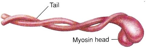
Figure 2.1 – a molecule of myosin
While some muscles may be arranged to act together to achieve work, often the muscles are arranged in pairs such that as one muscle contracts, the other muscle extends. This is known as flexion and extension, respectively. For example, during the flexion of the concentration curl, as the bicep contracts, the tricep extends. During the extension of the concentration curl, as the bicep extends, the tricep contracts.
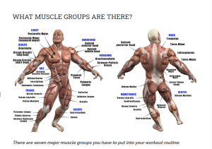
For each skeletal muscle, there is an origin and an insertion. The origin is the tendon connecting the muscle to the stationary bone, while the insertion is the tendon connecting the muscle to the mobile bone. During contraction, the muscle exerts a force on the mobile bone through the insertion tendon. Upper body muscles will be primarily be focused on for the purpose of this assignment. Refer to Figure 2.2 for a comprehensive diagram of the skeletal muscle system.
Muscle Growth
The growth of muscles is due to muscle fibre hypertrophy but it is thought that hyperplasia also plays a role in muscle growth. Muscle hyperplasia is essentially the increased number of fibres in the muscle due to splitting of the cells whereas hypertrophy is the increase in size of the muscle due to the enlargement in the size of the fibres. There are two types of hypertrophy in the muscles; transient and chronic. The main difference between the two is that transient hypertrophy is the increase in apparent size of the muscle due to fluid accumulation in the intracellular spaces of the muscle fibres while chronic hypertrophy is the increase in actual size of the muscle due to hyperplasia and the thickening of the individual muscle fibres.
Generally the increase in size of muscle fibres is dependant on protein synthesis.
Once the muscle fibres sustain microtears, the process of muscle growth is induced. Ultimately what occurs is that protein synthesis is increased due to a variety of hormones.
Testosterone, a steroid hormone, enters the muscle cell by diffusing across the cell membrane, and works together with a hormone receptor within the cell which then prompts gene transcription and protein formation. Human growth hormones (hGH) are released from the anterior pituitary (a gland in the head), and triggers the production of insulin-like growth factors (IGF). IGF causes the muscle cell to increase its uptake of amino acids and glucose to form protein while hGH causes nuclear division in the muscle cell without triggering mitosis and hence there are more nuclei to more rapidly synthesize protein. Protein synthesis can also be initiated due to the release of insulin within the body. The release of insulin can be stimulated by certain amino acids. Thus, protein synthesis is increased largely due to these three hormones, and with a larger amount and a faster rate of synthesis, proteins can rebuild the muscle fibres thicker.
For further information regarding steroids such as testosterone, refer to section
VIII.
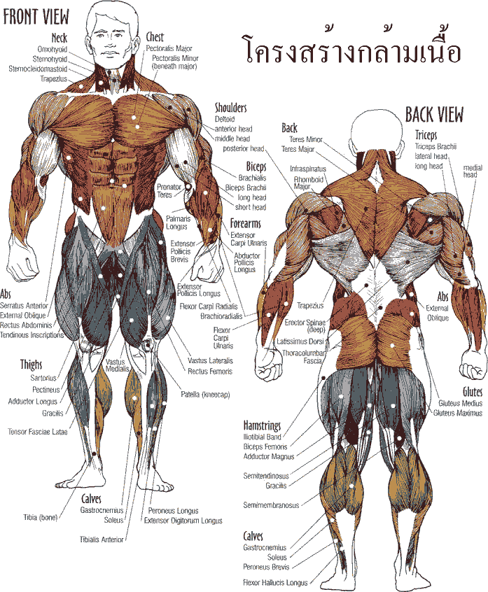
Figure 2.2-Muscle groups in the human body
Physical and Psychological Benefits of Exercising
There are numerous ways that exercising can be beneficial in both physical and psychological aspects of life.
Some of the more obvious physical benefits of exercising include less chance of death related to heart diseases and stroke, weight loss, get in shape and longer life span. Inactivity is one of the worst causes of heart diseases, along with smoking, high cholesterol levels, and high blood pressure. Exercise could help if not alleviate some heart problems present. As the heart is used with increased intensity, the cardiac muscle gets stronger, more efficient, pumping more blood per contraction, and thus puts less strain on the body. A recommended thirty minute medium-intensity exercise on most days of the week will reduce the risk of coronary diseases significantly. People who are active have a forty-five percent less chance of developing heart disease. A study done in 2002 reconfirmed the fact that even diminutive amounts of intense exercise could lower the cholesterol levels, while more exercise significantly lowers cholesterol levels.
Frequent exercise keep the arteries flexible, which improves blood flow and therefore keeps a regular blood pressure. Men who exercise merely one hour per day and five days per week, has a fifty gain percent less chance of getting a stroke.
Lungs also benefit from regular exercise. People with mild asthma gain from exercise by obtaining a larger breathing capacity. Even though 40% – 90% of asthma attacks are caused by exercising, bodybuilding exercises such as swimming, indoor running, and yoga are nevertheless good for asthma patients. A study showed that 66% of asthma patients who do yoga were successful in minimizing or eliminating the need for medicine.
For strong muscles and bones, exercise is important. As individuals age, muscles become less, but people who exercise are stronger and fitter than other people in their age group. A 2003 study proves that running could be associated with a longer life and less disabilities.
Exercising falls under 2 categories: strength training and cardiovascular training. The main goal of strength training is to build muscle mass while cardiovascular exercise achieves greater stamina by using light weights. Muscles build when they are put under strain. Exercising the main muscles groups for forty minutes 3-4 times a week is enough to increase muscle strength. Strength and massive muscles could be gained by using heavy weights and doing fewer repetitions. If the following symptoms are encountered, then overworking is possibly the cause: dizziness, constant fatigue, and frequent cold or the flu. Some of the positive effects of strength training would be: increasing muscle strength, increasing muscle size, more flexibility, balance, makes the bones stronger and denser, reduce risk of degrading diseases, help reduce fat levels, increases quality of life, and increasing the testosterone level. Cardiovascular training, on the other hand, is one where an activity is usually done over a longer period of time. It includes walking, jogging, swimming, and many other bodybuilding exercises. Cardiovascular training helps condition the heart and the lungs, ensuring a healthy and long life. Some other benefits of cardiovascular exercises include: a lean body, a strengthened heart, better cholesterol
(more good vs. bad), reduces chances of getting colon cancer, reduced risk of getting diabetes, and a lower heart rate and blood pressure, and a enhanced quality of life.
Psychological Benefits of exercising is essentially the mechanism behind how exercises help the exerciser psychologically is still a mystery, but is does have the following positive impacts on the exerciser: relieve stress and tension, improves self- esteem, boost blood flow to the brain, and just feeling better about yourself. Physical exercise acts like a buffer against stress by minimizing the effect of stress on the body. Regular exercise also decreases anxiety by stabilizing it. As the workout/exercise is begun, anxiety actually rises, but levels off as the activity is continued. Five to thirty minutes after the workout has concluded, the anxiety level is lower than before. It has shown a greater impact on reducing the effects of anxiety than the drug meprobamate, a powder that is used to relax muscles, relieves anxiety as a tranquilizer, and an anticonvulsant. Also it has a vastly constructive impact on the human body by relaxation and lowering anxiety, the effects are relatively short-term compared to certain drugs; the relaxation obtained from exercising diminishes in about 4 hours, and anxiety levels return to normal in 24 hours. Daily exercise is required if the person is suffering from chronic anxiety. However, overworking could be stressful and bad for both body and mind.
Tests have shown that a constant level of intensity of aerobic exercises enhances the brain’s capacity to process information the best. A study using soccer players showed that after running on a treadmill for 2- 45 minute time periods, their response time to a test involving real time soccer plays decreased with more time on the treadmill. In another separate study, female runners got better at solving simple math problems after a 20 minute run, and improved more after a 40-minute run. The mechanism behind the effect is not clear, but scholars believe that aerobic exercises act like stimulation drugs by releasing hormones and some chemicals such as adrenaline. However, working over the personal limit is not good for health.
Weight Training: Anaerobic Exercise Mechanics & Impact on Muscle Growth
-
- Changes of energy in exercising
- Motion in exercising
- Muscles acting as levers
- Investigate torque in bodybuilding
- Intensity vs. speed
Part A – Work & Energy transformations occurring during an exercise
During exercising, energy is supplied to the muscles as chemical energy. This energy is then converted to mechanical work (potential and kinetic energies) during the physical process of weight training. The ultimate objective of this particular lab is to determine the kinetic and potential energies of the weight (e.g. dumbbell) by using the equations Ek = ½mv2 and Ep= mgh.
For isotonic exercises, more mechanical work done means that more tearing of the muscles is occurring. By tearing the muscles, the muscle fibres will be mended, and made thicker than before.
Materials
-
- Meter stick
- Timer
- Free Weights (dumbbells, between 20 and 30 lbs)
- Human subject
Procedure
- The subject should begin doing a bench press exercise, in which he will pause at three stages of the exercise. These stages will include initial and final stages, as well as an “in-between” stage (refer to Figure 2.1)
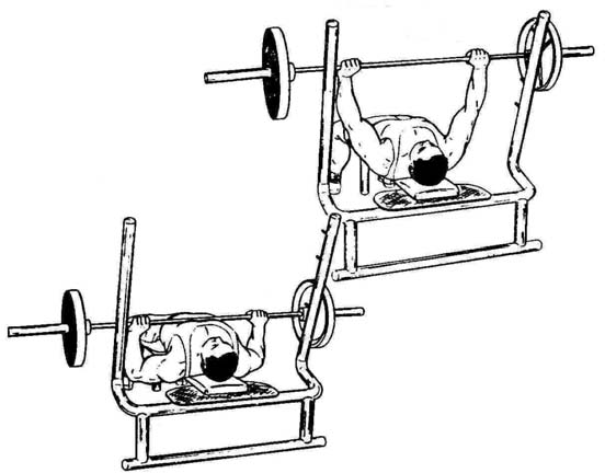
Figure 2.1- Initial and final stages of a bench press (using a barbell)
- Measure the displacement of the weight from the reference point, where Ep= 0, which is the lowest point during the exercise
- Measure the time to complete half a repetition (“rep”)
Observations
Table 1.1- Distance versus Time for half a repetition using 38.5lbs total
| Displacement of half a repetition, d (cm) | Time to complete half a repetition, t (s) |
| 46.50 | 1.06 |
Table 1.2- Distance versus Time for half a repetition using 43.5lbs total
| Displacement of half a repetition, d (cm) | Time to complete half a repetition, t (s) |
| 46.50 | 1.17 |
Table 1.3- Distance versus Time for half a repetition using 48.5lbs
| Displacement of half a repetition, d (cm) | Time to complete half a repetition, t (s) |
| 46.50 | 2.16 |
Analysis
Firstly, because our masses are in lbs, we must convert them to kilograms. 1kg = 2.2lbs, thus
-
- 38.5lbs/2.2 = 17.5kg
- 43.5lbs/2.2 = 19.8kg
- 48.5lbs/2.2 = 22.0kg
To calculate the velocity of each of the trials, we use the formula v = d/t
-
- for 38.5lbs: v = 0.4650m/1.06s = 0.439m/s
- for 43.5lbs: v = 0.4650m/1.17s = 0.397m/s
- for 48.5lbs: v = 0.4650m/2.16s = 0.215m/s
We will first calculate the amount of work done for half of a repetition using various masses. The formula W = F•d
-
- for 38.5lbs: W = (17.5kg * 9.81m/s2) * 0.4650m = 79.8J
- for 43.5lbs: W = (19.8kg * 9.81m/s2) * 0.4650m = 90.3J
- for 48.5lbs: W = (22.0kg * 9.81m/s2) * 0.4650m = 100J This is assuming the barbell is traveling at constant velocity.
To calculate the kinetic energy, the formula Ek = ½mv2
-
- for 38.5lbs: Ek = ½(17.5kg)(0.439…m/s)2 = 1.69J
- for 43.5lbs: Ek = ½(19.8kg)(0.397…m/s)2 = 1.91J
- for 48.5lbs: Ek = ½(22.0kg)(0.215…m/s)2 = 2.12J
To calculate the potential energy at the top of the extension of the exercise, the formula Ep = mgh
-
- for 38.5lbs: Ep = (17.5kg)(9.81m/s2)(0.4650m) = 79.8J
- for 43.5lbs: Ep = (19.8kg)(9.81m/s2)(0.4650m) = 90.3J
- for 48.5lbs: Ep = (22.0kg)(9.81m/s2)(0.4650m) = 100J
Discussion
For most isotonic exercises, the more work done by the individual in the process of bodybuilding, the better the workout is. This is due to the fact that generally muscles undergo more micro-tears when doing more work, which in turn signals for an increased production of actin and myosin filaments. However, in order to achieve maximum results during workouts, one must find the balance between the number of repetitions they do and the amount of weight they lift. If an individual attempts to weight train with weights that are too heavy, they won’t get a very good workout. This is because while they are doing more work per repetition, they are not capable of doing very many repetitions, and therefore don’t do very much work. On the other hand, a person working out with an overly light weight will be able to do a lot of repetitions, but the amount of work done per repetition will be miniscule. Furthermore, the human body experiences maximum muscle growth when skeletal muscles are put under a lot of stress and experience tears (accomplished by lifting heavy weights). Therefore, lifting light weights in your workout routine is not recommended for bodybuilding.
Conclusion
The quantity of work done during an exercise routine is a better measure of the energy expended rather than how much the routine affects muscle growth. However, within a reasonable intensity range, exercise routines in which a greater amount of work is done is usually better for bodybuilding purposes.
Part B – Investigating torque in weight training
Torque is essentially the rotational effect on a body due to an applied force. As an exercise involving the arm(s) is being performed, there is for a tendency for the arm to rotate. Thus, there is torque in the arm as it is being exercised. To calculate the amount of torque used, we use the formula τ = r┴ x F, where r┴ = r · sin θ. We can measure r┴ and F, as illustrated in the following diagram.
Excessive stress on the joints causes bone degradation, and can cause osteoporosis. The more weight you lift during workouts, the more torque your arm has during various phases of the exercise, and the more stress is put on your joints.
Therefore, people with bone problems should be conscious of how much weight they lift during workout.

θ
r
Figure 3.1- where r and θ occur during a bicep curl exercise
Materials
- Protractor
- Ruler
- Human subject
- Dumbbells (between ten and thirty pounds)
Procedure
- Record the length of the fulcrum to the load (r)
- The subject should begin a bicep curl pausing at five stages, including the initial and final stages (as demonstrated in Part A), while recording theta (the angle from the forearm to just below the bicep muscle) for each stage.
Observations
r is 32.50 cm
Table 2.1- The angles formed between the fulcrum and the lever arm at various positions in a bicep curl exercise using 10.0lb free weights
| Stage/Position in the bicep curl | Angle between upper arm and forearm, θ (o) |
| 1 | 180.0 |
| 2 | 140.0 |
| 3 | 90.0 |
| 4 | 60.0 |
| 5 | 20.0 |
Table 2.2- The angles formed between the fulcrum and the lever arm at various positions in a bicep curl exercise using 20.0lb free weights
| Stage/Position in the bicep curl | Angle between upper arm and forearm, θ (o) |
| 1 | 180.0 |
| 2 | 140.0 |
| 3 | 90.0 |
| 4 | 60.0 |
| 5 | 20.0 |
Table 2.3- The angles formed between the fulcrum and the lever arm at various positions in a bicep curl exercise using 30.0lb free weights
| Stage/Position in the bicep curl | Angle between upper arm and forearm, θ (o) |
| 1 | 180.0 |
| 2 | 140.0 |
| 3 | 90.0 |
| 4 | 60.0 |
| 5 | 20.0 |
Analysis
Table 3.4 – Torque of the dumbbell versus the force that needs to be exerted by the elbow joint at various positions in a bicep curl exercise using 10.0 lb free weights
| Stage/Position in the bicep curl | Torque of dumbbell,
τ (N·m) |
Force exerted by the bicep, FA, (N) | Force exerted by the elbow joint, Fs (N) |
| 1 | Negligible | Negligible | Negligible |
| 2 | 9.32 | 328 | 284
83.7° down from forward horizontal |
| 3 | 14.5 | 294 | 250
78.9° down from forward horizontal |
| 4 | 12.6 | 270 | 226
79.3° down from forward horizontal |
| 5 | 4.96 | 245 | 201
85.3° down from forward horizontal |
Table 3.5 – Torque of the dumbbell versus the force that needs to be exerted by the elbow joint at various positions in a bicep curl exercise using 20.0 lb free weights
| Stage/Position in the bicep curl | Torque of dumbbell,
τ (N·m) |
Force exerted by the bicep, FA, (N) | Force exerted by the elbow joint, Fs (N) |
| 1 | Negligible | Negligible | Negligible |
| 2 | 18.6 | 656 | 567
83.7° down from forward horizontal |
| 3 | 29.0 | 588 | 500
78.9° down from forward horizontal |
| 4 | 25.1 | 538 | 450
79.3° down from forward horizontal |
| 5 | 9.91 | 490 | 401
85.3° down from forward horizontal |
Table 3.6 – Torque of the dumbbell versus the force that needs to be exerted by the elbow joint at various positions in a bicep curl exercise using 30.0 lb free weights
| Stage/Position in the bicep curl | Torque of dumbbell,
τ (N·m) |
Force exerted by the bicep, FA, (N) | Force exerted by the elbow joint, Fs (N) |
| 1 | Negligible | Negligible | Negligible |
| 2 | 27.9 | 983 | 850
83.7° down from forward horizontal |
| 3 | 43.5 | 882 | 750
78.9° down from forward horizontal |
| 4 | 37.7 | 808 | 676
79.3° down from forward horizontal |
| 5 | 14.9 | 737 | 603
85.3° down from forward horizontal |
Sample Calculations:
Torque of Dumbell:
τ = r┴ x F = rsinθ x F
τ=(30.0lb/2.2)(9.81m/s2)(0.3250m)sin(180.0-90.0)=43.5 N·m
Force that needs to be exerted by the elbow joint:
First, we have to find θ for the applied force of the bicep. c2=a2+b2-2abcos(C)
We know that the bicep insertion (where the force of the bicep is applied) is approximately 5.00cm or 0.0500m from the fulcrum (the elbow). We also know that the distance between the elbow joint and the elbow is 0.3000m.
c2=(0.0500m)2+(0.3000m)2-2(0.0500m)(0.3000m)cos(90.0) c=0.3041381265…m
By using the sine law:
a/sin(A)=c/sin(C)
0.0500m/sin(A)= 0.3041381265…m/sin(90.0) A=9.462322208°
b/sin(B)=c/sin(C)
0.3000m/sin(B)= 0.3041381265…m/sin(90.0) B=80.53767779°
To find θ:
θ=180.0°-80.53767779°=99.46232221…°
Since the dumbbell-arm lever system is in rotational and translational equilibrium, we know that ∑F=0 and ∑τ=0.
Let the load be τl and let the applied force be τA. τ1-τA=0 τ1=τA
τ1=FA x rAsinθA
(43.5 N·m)=FA x (0.0500m)sin(99.46232221…°)
FA=882.0005666…N
∑Fx=0
Fx – (882.0005666…N)cos(90.0°-9.462322208…°)=0 Fx = 145 N
∑Fy=0
Fy-(882.0005666…N)sin(90.0°-9.462322208…°)+(30lb/2.2)(9.81m/s2)=0 Fy = 736.2267…N
a2+b2=c2
(145 N)2+(736.2267…N)2=c2 c=750 N tanθ=(736.2267…N)/(145 N)
θ=78.9° down from forward horizontal
Discussion
Maximum torque is generated by the dumbbell at phase 3 of the exercise (where the forearm is parallel to the ground). This makes sense because when the force gravity provided by the dumbbell is perpendicular to the forearm of the individual doing the bicep curl, the lever arm will be at its greatest length. Therefore, since τ = r┴ x F where F is a constant, maximizing the length of the lever arm will yield in maximum torque generation. As the forearm shifts away from the maximum lever arm position, the torque generated by the load will decrease proportionally.
According to our lab results, as the angle between the upper arm and the forearm decreased, so did the stress on the elbow joint. However, according to various sources on the internet, maximum stress on the elbow joint should occur when the forearm of the individual doing the exercise is parallel to the ground. Internet sources also stated that the torque generated by the dumbbell in this exercise should be directly proportional to the amount of stress the elbow joint is put under. This discrepancy is most likely caused by some kind of experimental error. The lab results obtained in this lab show us that even by doing the bicep curl with fairly light weights, a large amount of force is exerted on the elbow joint. Using excessive weight during workouts or working out too often can lead to bone degradation, which causes many problems later on in life (including arthritis). Bodybuilders are advised from doing exercises that put excessive stress on joints in the body because more muscle mass at the expense of skeletal health is not worth it in the long run.
Conclusion
The force exerted by the bicep in the bicep curl is directly proportional to the amount of stress the elbow joint is put under. However, due to conflicting research and lab results, it’s not clear whether or not the amount of torque generated by the dumbbell is directly proportional to the amount of stress the elbow joint experiences. Further
experimentation is required before these lab results can be considered conclusive.
Part C – Muscles acting as levers
A lever is fundamentally a device which allows the movement of a load using a force around particular pivot point. Levers can be divided in to three different classes. Arms are generally third class levers, which simply signifies that there is a stationary pivot point and a load at the two extremes and an applied force in the middle. An example of this is the concentration curl where the elbow remains stationary while the forearm lifts the dumbbell, which represents the load. It is the bicep which generates the applied force but the force is transferred to the forearm by the bicep insertion, the tendon which connects the bicep to the forearm. The unique characteristic of this particular class of levers is that the muscle does not need to contract much although it inverse proportionally exerts a much greater force in order to create a great deal of movement of the load.
Because arms are generally third class levers, very little movement in the muscle causes a significant amount of movement of the load which means a greater amount of force is needed to move the load. Thus, the arm can move a load rapidly but is ineffective at lifting heavy loads relative to secondary and first class levers.
Due to the fact that humans the bicep-arm system is a third class lever. The muscle must exert a greater force than that provided by the load. Therefore, more muscle tearing occurs than if the arm is a 2nd or 1st class lever, which is beneficial for bodybuilding.
Figure 4.1-the arm acting as a third class lever
To demonstrate that the arm is quite ineffective at lifting heavy loads, a ratio between the contraction of the muscle and the movement of the load can be determined.
Materials
-
- Human subject
- Ruler
- Dumbbells (15lbs)
- Protractor
Procedure
- Measure the length between the fulcrum and the load
- The subject should begin a concentration curl pausing at the initial and final stages while recording the angle between the initial and final stages of the arm as demonstrated in the diagram below.
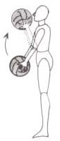
Figure 4.2-initial and final positions in a bicep curl
- Measure the contraction of the bicep muscle at the initial and final positions
Observations
Table 3.1-Displacement from the fulcrum to the bicep versus the angle of movement between initial and final positions of the arm
| Trial | Initial angle between forearm and upper arm, θi (o) | Final angle between forearm and upper arm, θi (o) | Displacement from fulcrum to bicep initially, di(cm) | Final Displacement from fulcrum to bicep, df (cm) |
| 1-Di | 130.0 | 40.0 | 1.10 | 7.00 |
| 2-Ajay | 130.0 | 40.0 | 1.50 | 7.50 |
-
- Di’s distance between the fulcrum and the load (dumbbell) = 32.50cm
- Ajay’s distance between the fulcrum and the load (dumbbell) = 34.20cm
Analysis
Because the motion of the arm is not straight, we must calculate the arc length of the displacement, using the formula arc length = r*. However, we must first convert degrees to radians:
(130.0o – 40.0o) * (π/180o) = 1.57rad
-
- arc length 1 = r* = (32.50cm) * 1.57rad = 51.1 cm
- arc length 2 = r* = (34.20cm) * 1.57rad = 53.7 cm
Now to calculate the ratio of how much the bicep moved versus how much the load moved:
Displacement of load/displacement of muscle=(51.1cm)/(7.00cm-1.10cm)=8.66 Displacement of load/displacement of muscle=(53.7 cm)/(7.50cm-1.50cm)=8.95 Average=(8.66cm+8.95cm)/2=8.81cm
On average, for every centimeter the bicep contracted, the load moved 8.81 cm.
Conclusion
According to the formula for torque (τ = r┴ x F) and our lab results, the bicep must exert a force 8.81 times as big as the load because the load arm is approximately 8.81 times as big as the force arm. This is advantageous during workout because you can get the bicep to exert a comparatively large force on a load that may not weigh very much.
More muscle filaments are torn when the bicep must exert a massive force to lift the load and therefore triggers the production of bulkier actin and myosin filaments. Bulkier actin and myosin filaments make the muscle bigger and stronger than before, which is what bodybuilding is all about.
Part D – Impulse in weight training
During an exercise, at the halfway point of a repetition, it can often be disadvantageous to let the weight drop. For example, during a bench press exercise if the barbell is dropped, the weight will simply accelerate down. The barbell must be stopped with a great amount of force with the sternum, various ligaments & tendons applying an opposing force and the barbell must be stopped in a very short period of time before it falls on the subject’s neck. Thus in this scenario, the pectorals are not getting the maximum amount of workout as possible relative to letting the weight down with a constant velocity. Thus, when the barbell is kept at a constant velocity on the way down, the force applied is less because it is over a greater amount of time. The equation to determine the impulse is impulse=Fnet · ▲t and thus when a constant velocity is kept, Fnet=0 and the impulse is equal to zero. Hence, when the impulse is zero, the exercise is generally more effective.
 Letting the weight drop during the bench press is very harmful to the tendons in the shoulder and chest region. For this situation, Fnet∆t=m∆V, where m∆V is a constant. The force that is exerted on the tendons in the shoulder and chest region is increased dramatically because the barbell is being stopped in a short amount of time (before the barbell can kill the person). This causes tendon damage in these areas.
Figure 5.1-initial and final positions of a bench press exercise using a barbell Materials
- Stopwatch
- Human subjects
- Dumbbells (5lbs)
- Ruler
- Elastic
Procedure
- Have the subject lift the free weights until the arms are fully extended
- The subject should let the two dumbbells drop but stop them before they hit the sternum, as shown in the final position of Figure 5.1
- Have the subject lift the free weights until the arms are fully extended
- Measure the distance between the load and the parallel of the sternum
- The subject should then bring the weights down with a relatively constant velocity
- Measure the length of the elastic at equilibrium position
- Attach a 5 pound weight onto the elastic
- Displace the mass upwards from equilibrium position by an indicated amount (5cm, 10cm, 15cm, 20cm, 25 cm)
- Release the mass
- As soon the mass goes past the equilibrium position, start the timer
- Stop the timer when the mass stops
- Record results.
Observations
Table 5.1-distance between the load (10.0lbs) and the parallel of the sternum for various trials
| Trial number | Distance between the load and the parallel of the sternum, d (cm) |
| 1 | 48.33 |
| 2 | 53.25 |
| 3 | 46.50 |
Table 5.2-diplacement of the mass from the equilibrium position of the elastic versus the time it takes for the elastic to stop the mass
| Displacement of the mass from the equilibrium position of the elastic, d (cm) | Time taken for the elastic to stop the mass, t (s) |
| 15.00 | 0.5800 |
| 20.00 | 0.5600 |
| 25.00 | 0.5300 |
| 30.00 | 0.3900 |
| 35.00 | 0.4405 |
Analysis
Table 5.3-Velocity of mass just before an opposing force is applied on it versus the average magnitude of the opposing force
| Velocity of mass just before an opposing force is applied on it, v (m/s) | Average magnitude of opposing force, Fav (N) |
| 1.72 | 52.4 |
| 1.98 | 60.7 |
| 2.21 | 63.6 |
| 2.43 | 72.9 |
| 2.62 | 71.6 |
Sample Calculation:
Velocity of mass just before an opposing force is applied on it: Vf 2 = Vi 2 + 2ad
Vf 2 = 0 + 2(9.81 m/s2)(15.00 cm/100)
Vf =1.72 m/s
Average magnitude of opposing force:
Fnet∆t=m∆V
Fnet(0.5800s)=(10.0 lb/2.2)(1.72 m/s) Fnet=7.82 N
Fav=7.82 N + (9.81 m/s2)(10.0 lb/2.2)=52.4 N
Discussion
Tendons are actually quite elastic, much like the elastic bodies used in this lab.
From the results of this lab, we can conclude that as the distance the mass was allowed to undergo free fall increased, the average force required to stop the mass also increased.
We can postulate that increasing the mass used in this lab will also have a similar effect on the average force required to stop the mass. According to Hooke’s Law (F=kx), as the maximum force required to stop the mass increases, the amount of “stretch” the elastic undergoes also increases. Therefore, by increasing the distance the mass is allowed to fall and/or by increasing the mass itself, we can say that the elastic will undergo a greater degree of stretch. The tendons in the human body, much like the elastics used in this experiment, have a certain elastic limit. Once it’s stretched beyond this limit, it will become permanently deformed and may even break. This causes various problems, because tendons have various roles in the muscular system of the body, including saving energy during workouts and improving muscular control. Because tendons are what connect skeletal muscles to the skeleton, caution should be exercised to not damage the tendons in your body during workouts, or you will find yourself not being able to do everyday tasks very well.
Another drawback to using bad technique like dropping the weight on oneself during the bench press exercise is that the tendons are what absorb most of the shock from stopping the barbell before it kills the individual, so the pectoral and shoulder muscles don’t get a very good workout. Maximum muscle stress is achieved by keeping the barbell at constant velocity during the extension and flexion phases of the exercise, in which case the impulse of the barbell should equal 0.
Conclusion
Proper technique should be followed in bodybuilding to prevent tendon injuries. Bodybuilding is about improving one’s physical condition, not worsening it.
Part E – Influence of speed and intensity of workout on blood pressure and heart rate
A faster and more intense workout will yield a higher blood pressure and heart rate in the human subject.
The heart is described as “hollow muscular organ”; its function is to pump blood to the whole body during a person’s life. The circulatory system of which the heart is part of sends oxygen and nutrients to the body and removes waste and carbon dioxide. One of the demands on the heart is that it must be able to shift whenever the activity of the person changes. Thus when activity increases and a person were to exercise, they would need more oxygen. The increase in oxygen demand leads to the heart being forced to pump more blood and therefore the heart rate of that particular individual to increase. “Heart rate” simply signifies the number of beats the heart undergoes per minute. Under normal circumstances, the heart rate of and adult would be seventy beats per minute while that of a child would be one-hundred beats per minute and a baby’s would be one- hundred and twenty beats per minute. Resting heart rates will increase due to exercise training. For example, professional athletes have slower resting heart rates due to physical training which keeps the heart stronger so that it may pump a higher volume of blood while beating less often. Other sources such as stress, temperature, hormones, drugs, alcohol and food can also affect heart rate.
Blood pressure is essentially the force of one’s blood on the arteries’ walls. It is measured by the systole, the “highpoint” where the heart releases blood by contracting, and the diastole – the “low point” in which the heart relaxes and thus is filled with blood. When blood pressure is measured, it is generally measured in mm of mercury (mmHg) in a sphygmomanometer. The maximum blood pressure is systole and the minimum is the diastole for a “cardiac cycle”. Normal blood pressure should be should be 80/45 in babies while individuals at thirty years of age should have a blood pressure of 128/80. If ones blood pressure were too high, some of the blood vessels could explode, yet if the blood pressure was too low, the brain would “starve” or not be able to get what it needs. Thus, the body controls blood pressure in various ways to meet its needs. For example, it can tighten or loosen the blood vessels also the heart can change the amount of blood it pumps. Blood pressure will be affected by the flexibility of arteries, the diameter or width of the artery, the thickness or viscosity of blood and the volume of blood. The volume of blood can change if a lot of blood is lost causing the blood pressure to go down. As well, the heart rate can affect blood pressure. As one exercises, their heart rate increases thus resulting in an increase of blood pressure. As the heart rate decreases, the blood pressure decreases.
Materials
-
- Blood pressure monitor
- Three humans
- Stop watch
- Ruler
- Barbell
- 6 stool and a cushion or a bench press
Procedure
- Set up stools and cushions refer so that the stools are in a line and some cushions and towels care padding it , use a light and small cushion for the head and a few towels for the rest if wanted
- Have the human subject sit relaxing and not really doing much for at least roughly 10 minutes
- Measure the subject’s blood pressure and heart rate
- Have the subject start on the bench press
- Measure the distance travelled by the barbell from the bottom to the top and the time it takes for the barbell to travel this distance
- After the exercise, measure the subject’s blood pressure and heart rate
- Repeat the steps for the wanted trials
Observations
Table 6.1 – Heart rate and blood pressure before and after an intense workout (8 reps) using 58.5lbs
| Trial # | Heart rate before workout, h (beats/min) | Blood pressure before workout, p (mm hg) | Heart rate after, h’ (beats/min) | Blood pressure after workout p’ (mm hg) | Distance travelled by weight for half a repetition, d (cm) | Time taken for half a repetition, t (s) |
| 1-Di | 78 | 118/89 | 89 | 120/80 | 48.33 | 1.85 |
| 2-Kelei | 101 | 110/63 | 116 | 109/82 | 46.50 | 2.06 |
Table 6.2 – Heart rate and blood pressure before and after a fast workout (20 reps) using 23.5lbs
| Trial # | Heart rate before workout, h (beats/min) | Blood pressure before workout, p (mm hg) | Heart rate after, h’ (beats/min) | Blood pressure after workout, p’ (mm hg) | Distance travelled by weight for half a repetition, d (cm) | Time taken for half a repetition, t (s) |
| 1-Di | 78 | 118/89 | 93 | 134/73 | 48.33 | 0.85 |
| 2-Kelei | 101 | 110/63 | 120 | 104/89 | 46.50 | 0.81 |
Analysis
Table 6.3 – Velocity of the mass during the workout versus the change in heart rate and blood pressure after an intense bench press workout (8 reps using 58.5 lb)
| Trial # | Velocity of mass, v (m/s) | Change in heart rate, ∆h (beats/min) | Change in blood pressure, ∆p (mm hg) |
| 1-Di | 0.261 | 11 | 2/-9 |
| 2-Kelei | 0.226 | 15 | -1/19 |
Table 6.3 – Velocity of the mass during the workout versus the change in pulse and blood pressure after an fast bench press workout (20 reps using 23.5 lb)
| Trial # | Velocity of mass, v (m/s) | Change in heart rate, ∆h (beats/min) | Change in blood pressure, ∆p (mm hg) |
| 1-Di | 0.57 | 15 | 16/-16 |
| 2-Kelei | 0.57 | 19 | -6/26 |
Discussion
In part B of the lab section, we investigated how the quantity of work done during bodybuilding exercise routines affects the effectiveness of these routines. However, the amount of work done during a workout is just one of the factors that influence how effective the workout is. This is shown very clearly by the results of this lab. Two individuals executed an intense workout, followed by a rest period, then a fast workout. The amount of work done in both exercises is about the same. However, both individuals clearly experienced differences in how much their heart rates and blood pressure changed during the two workouts. The fast workout seems to yield a greater change in heart rate and blood pressure. Why did this occur? Our hypothesis is that during the intense bench press exercise, the demand for ATP was so high that the body must resort to anaerobic respiration to supply the necessary muscles with energy. Anaerobic respiration involves glucose getting turned into pyruvate, then being converted to lactic acid, and finally it’s converted back to glucose in the liver and the process continues. Anaerobic respiration produces ATP, but does not create CO2 as a waste product. The body does not need to get rid of an increased quantity of CO2, so breathing rate, heart rate, and blood pressure all remain the same. However, anaerobic respiration and aerobic respiration must occur and the same time to supply the skeletal muscles with enough ATP. Since aerobic respiration does produce CO2 as a waste product, breathing rate, heart rate, and blood pressure will go up dramatically. The fast bench press workout relied on mainly aerobic respiration while the intense bench press workout relied on both aerobic and anaerobic respiration, therefore the individuals who did the fast workout experienced a greater change in blood pressure and heart rate. The same individuals doing the intense workout experienced a lesser change in blood pressure and heart rate
Conclusion
While it seems that the change in heart rate and blood pressure of the two individuals executing the bench press exercise is influenced by the velocity at which they are moving the barbell, more scientific testing is needed to confirm this. The results of this lab indicated that fast bench press workouts results in a greater in heart rate and blood pressure than the intense bench press workouts. Therefore, for general health purposes, fast workouts are better because it gets heart rate up more. Increased heart rate during exercise trains the heart to pump more blood per contraction, which increases the fitness level of the individual. For bodybuilding purposes, the intense workout is better because muscle filaments are being re-synthesized stronger and bulkier than before.
Research indicates that while aerobic exercise results in a higher endurance of the muscle, the size of the muscle itself does not change.
Protein Supplementation
Protein-powder is a very popular supplement, and is available at most local drug and health food stores. Before proteins can be used for muscle hypertrophy in the body, they must be broken down to amino acids in the liver. In the liver, they can be re- synthesized into muscle proteins, and transported to the muscles for hypertrophy.
Obviously, if the proper stimulus is given, the more amino acids responsible for muscle growth that are present in the body, the more muscle hypertrophy will occur. Simply put, the more protein in the body, the more muscle hypertrophy will occur.
Studies show that ingestion of protein with carbohydrate increases insulin and/or growth hormone level, thus assisting the process of muscle growth. The intake of protein and carbohydrate before exercise boost the efficiency of the workout and lessens recovery time, allowing the athlete to endure more vigorous training sessions. As much as 1.3 to 1.8 g of protein for every Kg of body mass, or 2.2 g at high altitudes, is recommended for weight trainers per day. It is shown that 20 calories per pound of body mass is required to sustain muscle mass, and 25 to 30 calories per pound to actually build muscle mass. Thirteen to seventeen percent of those calories should come from proteins, with twelve to sixteen percent coming from fats, and the rest obtained from carbohydrates.
Tests show that taking protein supplements along with creatine monohydrate works better than just taking protein supplements alone. The positive effects include leaner tissue mass, and increase in bench press power. Another independent study shows how a combination of protein and creatine helps boost muscle GLUT-4 (glucose transport proteins) matter and increases glucose tolerance in the athlete. Human subjects’ right leg was immobilized for the time period of two weeks, and then resistance exercises were put in place for six weeks. GLUT-4 content was decreased in creatine-taking subjects, creatine and protein-taking subjects, and placebo-taking subjects during the 2 weeks of immobilization. When the resistance exercise program was introduced, the GLUT-4 content in creatine-taking subjects were up by 24%, creatine and protein-taking subjects’ GLUT-4 content were increased by 33%, while placebo-taking subjects’ GLUT- content remained constant. Muscle glycogen content in both creaine-taking and creatine and protein-taking subjects, but not placebo-taking subjects.
Reports show that carbohydrate intake along with protein supplementation increases insulin and/or growth hormone levels a relatively large amount. As well, intake of carbohydrates and protein subsequent to anaerobic exercise promotes a “more anabolic hormonal profile”, as well as glycogen (which is discussed in the following section) resynthesis, and potentially faster muscle recovery.
Cellular Respiration & Effect on Weight Training
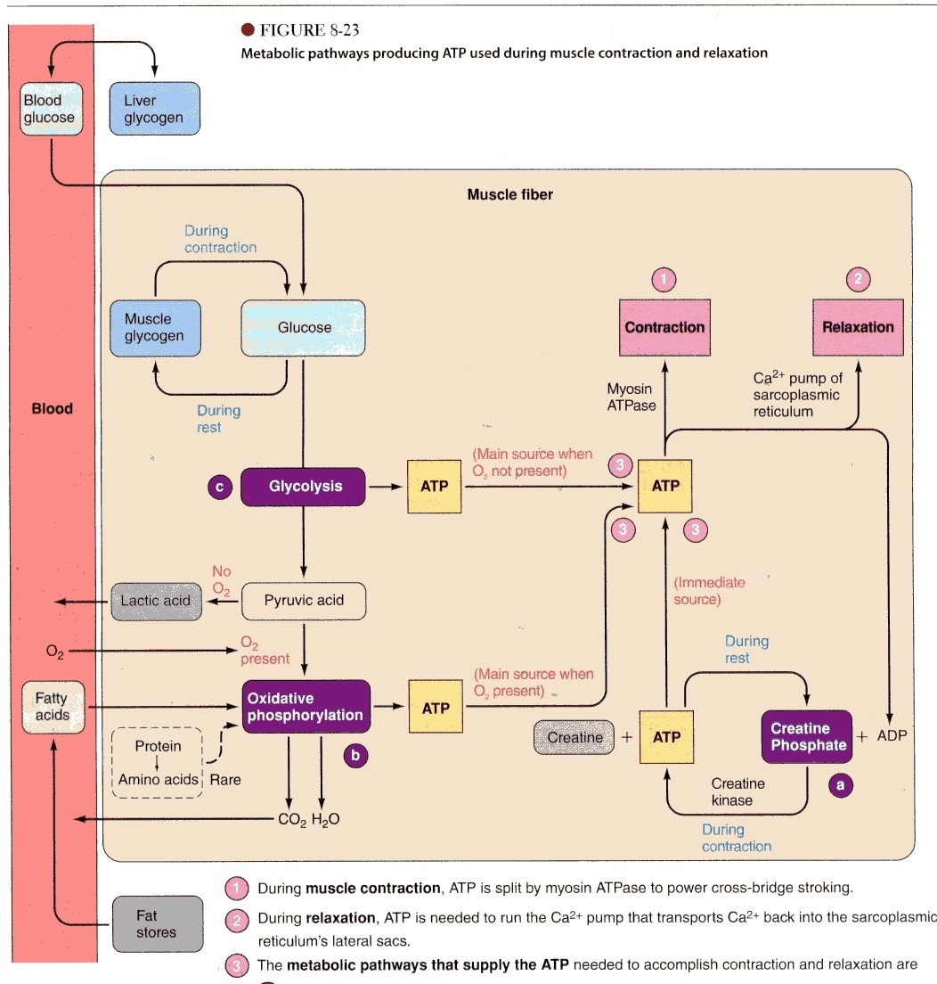
Figure 5.1 – metabolic pathways producing ATP used during muscle contraction and relaxation (copyright property of Human Physiology: from cells to systems). a)production of ATP supplied by the dephosphorylation of creatine phosphate, catalyzed by the creatine kinase. b)oxidative phosphorylation is the main source of ATP production
when oxygen is present (aerobic respiration). c)glycolysis is the main source of ATP production when oxygen is not present (anaerobic).
Glycolysis
Glycolysis is the beginning of a process in which the body converts glucose into energy. The body’s energy “currency” is called adenosine triphosphate (ATP). The process begins when there is glucose (a molecule consisting of 6 oxygen, carbon and hydrogen atoms) in the cytoplasm. In the cytoplasm, the glucose molecule is capped on both sides with a phosphate (a phosphate molecule consists of a phosphorus atom and four oxygen atoms) molecule. This is a type of phosphorylation (phosphate is added to a molecule) in which phosphate groups were added. This accomplishes two things for the glucose; it causes it to be more reactive, and become trapped in the cell. Subsequently, an enzyme divides the glucose molecule in half yielding two molecules each with its own phosphate group. At this time, two hydrogen atoms will move off each of these two molecules. The hydrogen is then placed on NAD+ (nicotinamide adenine dinucleotide) which yields NADH. While this occurs, the two molecules that were a result of glucose being divided have a phosphate molecule added to each. This essentially makes two reactive molecules which will twist itself into a different form, removing its phosphate groups to make a total of four ATP from adenosine diphosphate. Now, the molecules are made of three oxygen, three carbon and three hydrogen atoms and are called pyruvate or pyruvic acid. Essentially,
Glucose + 2 ADP + 2 NAD+ + 2 Pi → 2 Pyruvate + 2 ATP + 2 NADH + 2 H+
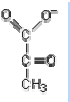
Figure 5.1 – structure of pyruvate, C3H3O3
In the case that there is oxygen present (aerobic), the transition stage, the Krebs cycle and finally the oxidative phosphorylation are undergone. If there is no oxygen present, the pyruvate is converted to lactic acid through the process of fermentation.
The Transition Stage
The transition stage is actually a link between glycolysis and the Krebs cycle. The pyruvic acid molecules are placed into the mitochondria where there is a folded membrane containing enzymes dictating the further steps of cellular respiration. Carbon dioxide, CO2, is removed from each pyruviate molecule; then hydrogen atoms and electrons from the pyruvic acid are added to two NAD+ hydrogen carriers, making two NADH. Once this has occurred, an enzyme takes one carbon atom and two oxygen atoms from each pyruvic acid molecule and thus the pyruvic acid molecule become acetyl groups. The acetyl groups are then connected to two coenzyme A yielding two molecules of acetyl coenzyme A.
Krebs Cycle
The third part of cellular respiration is called the Krebs Cycle. Here the acetyl coenzyme A molecule in the mitochondrial is connected to the oxaloacetate along with water to form citric acid. Subsequently, the acetyl coenzyme A is taken from the citric acid piece by piece as carbon dioxide and hydrogen groups and the result is an oxaloacetate, which can then take another acetyl molecule and repeat the same steps.
Two hydrogen atoms that are taken from the acetyl coenzyme A are placed on FAD++ (falvin adenine dinucleotide), yielding FADH2 while the other six are taken by NAD+. There are is only one ATP produced from every acetyl CoA molecule that enters the Kreb’s Cycle. The ATP is formed when coenzyme A is released in an exergonic reaction. Most of the ATP that are produced in cellular respiration will be produced in the next stage of cellular respiration, known as oxidative phosphorylation.
Oxidative Phosphorylation
In the electron transport chain, the NADH and FADH2 have carried the hydrogen atoms and its electrons so that they can divide them, thus resulting in protons and electrons. The electrons are placed close to the “electron transport chain molecules”, which consists of molecules that attract electrons and take them, following the first molecule the next molecule will attract the electrons even more and thus will take the electron away form it. After that the following molecule will take the electron away, this continues in the inner membrane of the mitochondria otherwise known as the cristae.
Each molecule has a stronger attraction for the electrons than the last one and thus is able to pull the electron away from its predecessor (these electron accepting molecules are referred to as the cytochrome carrier system). During this transportation of electrons, hydrogen ions are pulled from the matrix into the intercellular space. This chain will keep moving till the electron reaches oxygen, where oxygen will accept the electrons and thus become negatively charged and consequently can attract the hydrogen ions.
Hydrogen ions are pulled across the cristae from the intercellular space back into the matrix due to this electrostatic attraction and the fact that there is a concentration gradient of H+ ions between the intercellular space and the matrix. The hydrogen ions re-entering the matrix must pass through something called the ATP synthase. The ATP synthase uses energy this proton motive force to synthesize ATP molecules from ADP and phosphate ions. This process of oxidative phosphorylation makes a total of 34 ATP, water and causes the remaining NAD+ and FAD++ to be used.
In total, the process of cellular respiration yields 36 ATP using a single glucose molecule, two net are form gycolysis another two were made in the Kreb’s cycle and finally 32 were made in oxidative phosphorylation. This entire process is called aerobic respiration. In the case that there is no oxygen, aerobic respiration is undergone, where there is fermentation which follows the glycolysis, as mentioned previously.
Fermentation
After glycolysis there is fermentation, although there are two possible types of fermentation: lactate and alcohol. In muscle cells, lactic acid is a product anaerobic respiration. Lactate is a term that refers to the salts made from lactic acid and it is not
uncommon that both of these words were used as equivalents. In this case, the pyruvate molecules that are a result of glycolysis accept hydrogen and electrons from the NADH, yielding more NAD+ – this also changes the pryuvate into lactate. This type of energy production is for times in which ATP is needed very quickly, and there is a great need for it. Lactic acid can also lead to muscle pains that occur due to exercise, and another result of lactic acid can be a heart attack. Finally, the lactic acid is removed by blood flow.
ATP and the Phosphagen Energy Cycle
The way in which ATP (adenosine triphosphate) is used for energy is due to its structure. ATP consists of three phosphate groups; when the third phosphate group is broken off from the rest of the molecule, energy is released by breaking its high energy bond. This results in ADP (adenosine diphosphate). During exercise this molecule is phosphroylated by phosphocreatine with the assistance of the enzyme creatine kinase, yielding ATP again for the body to use. Creatine kinase also helps to add the phosphate to phosphocreatine by removing phosphate form the phosphocreatine, thus it can be added to the ADP yielding ATP and creatine.
Usually the cell will have five times the phosphcreatine as it has ATP and when exercise begins, there is only sufficient ATP to begin muscular contractions; and so the phosphocreatine helps to quickly replenish the needed ATP for a short time. Yet later on, the body is forced to use other ways to form ATP. Another function of the phosphagen energy system is that it helps to partly buffer the lactic acid produced during anaerobic respiration thus preventing possible muscle pains. This is done when the phosphate is being transferred from the phosphocreatine to the ADP to make ATP “consume” the H+.
One must not confuse the type of muscle soreness due to lactic acid build up (acute muscle soreness) and “delayed onset muscle soreness” which is also referred to as DOMS. Acute muscle soreness is the soreness one may experience due to H+ build up that comes from the lactic acid, this sensation will occur within hours to minutes of the exercise. Whereas DOMS is caused by either “structural damage” which is due to the increase in enzymes in muscle after exercise, this may cause tissue breakage. Another possible cause of DOMS is the possibility of an increase of white blood cells in the body after exercise that may cause “inflammatory reactions”.
Carb-loading
The idea behind carbohydrate-loading (which many athletes use in anaerobic exercise) is that by intaking many carbohydrates, exhaustion (which is caused by the depletion of glycogen-the primary storage form of glucose) can be delayed. This delay of exhaustion is due to maximizing the amount glycogen storage, consequently elevating the amount of ATP formed through the process of glycolysis (note that the glucose monomer is removed from glycogen prior to glycolysis).
Creatine Supplementation
Introduction
Creatine (methylguanidine-acetic acid) was discovered in 1832 by Michel Eugene Cheverul. Later on, in 1834 Justus von Lieburg “confirmed” that creatine was a normal part of meat. It was also found that there was more creatine in wild animals which underwent more exercise than animals that were living in captivity which exercised less. During the early part of the 1900s by using creatine as a supplement allowed for a boost in creatine in animals. Later on, phosphocreatine (creatine phosphate or phosphorylated creatine) was discovered in the year of 1927. Then in 1934, the creatine kinase (the enzyme that “catalyzes” phosphocreatine was found). Finally, in 1968, phosphocreatine was found in the process of recuperating from exercise.
In foods, creatine is found primarily in red meat and fish. Eaten creatine is then eventually sent to the bloodstream. Creatine is also synthesized within the body by the liver, kidney and pancreas, although this primarily takes place in the liver. This is done in two steps: the first step is when an amidine group from arginine goes to glycine to make guanidinoacetic acid. Then in step two, a methyl group goes to a guanidinoacetic acid from S-adenoslymthionine forming creatine. In the synthesis of creatine, there are some controls on it so that when there is less creatine in one’s diet, there will be more synthesis of creatine in the body. In opposition, if there is a lot of creatine present in one’s diet, then there will be less creatine synthesis in the body.
The storage of creatine in the body occurs in two forms; in the form of phosphocreatine or simply creatine. In the average adult male weighing 70kg, there is 120g of creatine of which 95% is found in the skeletal muscle. Some of the creatine goes to other various parts of the body such as the heart and brain. Of all the creatine in the skeletal muscles, 60-70% of that creatine is phosphocreatine. And because it is phosphocreatine, it cannot leave the membranes.
Lab : Impact of Creatine Monohydate and Phosphocreatine on Lactic Acid Buildup
The purpose of this lab is essentially to assess the buffering capacity of creatine monohydrate and phosphocreatine. As well, these two supplements will be contrasted to determine which of the two acts as a better buffer for the H+ ions (if at all).
Among many claims of the benefits of creatine, it has been advertised to “buffer” the build-up of lactic acid in muscles, thus delaying the process of burn in the muscle. A biological buffer is essentially a mechanism within the body which neutralizes H+ ions, which are ultimately excreted. As discussed before, lactic acid is essentially formed during anaerobic gycolysis, which occurs in the body when there is a lack of oxygen.
After various steps of gycolysis (refer to section VI), fermentation will take place. There are two types of fermentation, but for the purpose of this lab, lactic acid fermentation will be focused on. What occurs is the pyruate molecules from gycolosis accept the hydrogen and electrons from NADH yielding NAD+. The pyruvic acid is made into lactic acid, a
compound consisting of three carbons. In the body, lactic acid is formed not long after the acid dissociates, and will form a salt with chemicals such as Na+ and K+. Note the term lacate refers to any salt that is made from lactic acid. As well, when lactic acid builds up it can then cause acute muscle cramps and if enough of the acid is built up, it can cause a heart attack. Lactic acid is also considered to be responsible for muscle fatigue, as mentioned above.
It has been shown in various studies that phosphocreatine acts as a buffer in muscles against the hydrogen ions of acids, and in this particular case, lactic acid. Adenosine triphosphate (ATP) is the body’s form of energy and is made through several metabolic pathways. The actual structure of this molecule consists of an adenosine, a ribose, and three phosphate groups. A phosphate group is made of a phosphorus atom, and three oxygen atoms bonded to it. When one of the phosphate groups bonds to the molecule, it is broken by and enzyme called ATPase, and energy is released. There is a remaining adenosine diphosphate (ADP) and an “inorganic” phosphate group. Creatine begins to play a role in this phosphagen system and is phosphorylated by the creatine kinase enzyme, consequently yielding phosphocreatine. During intense exercise, the phosphate group that was originally added onto the creatine is being removed and placed onto the ADP making it ATP for ready use. (please note for more information on this entire subject please refer to section VI).
Phosphocreatine is responsible for approximately thirty percent of the muscle’s capacity to buffer the H+. As well, phosphocreatine uses up the H+ ions during the phosphagen energy system when the ATP is re-synthesized by phosphocreatine and ADP. (for more information to these various subjects refer to section VI).
There are three types of buffers within the human body; protein buffers, carbonic acid-bicarbonate buffers, and phosphate ion buffers. Although claims have been made that creatine is responsible for the buffering of lactic acid, the buffering is likely due to the phosphate in the phosphorylated creatine. The role of phosphate buffers within the body is to buffer primarily the intracellular environment and urine, although it plays a small role in buffering extracellular fluid.
(Note that due to the lack of availability of relatively pure phosphocreatine, it was not possible to obtain it. Thus, the lab will be performed using a creatine supplement containing a small amount of phosphate in it. However, there are many other ingredients in the mix which may affect the outcome of the lab. While there appears to be no protein within the supplement, it is not certain, which could significantly alter the results. For example, in some supplements there is hydrolysed vegetable protein, which would definitely have an unwanted impact on the results. Nevertheless, for the purpose of this lab, the assumption that any buffering is due to the phosphate will be made.)
To determine the pH of the titration, a universal indicator will be used. This indicator is unique in the sense that it consists of a series of indicators combined and thus has a higher range or reading level. And each one of the mixture of indicators shall
change its color at a different pH. A red color indicates a pH of one, while green indicates seven and purple indicating thirteen.
Materials
-
- beakers x 3
- lactic acid
- Erlenmeyer flasks x 3
- white paper towels
- distilled water in squeeze bottles x 3
- pipettes and bulbs x 3
- burette with stopcocks x 3
- retorque stand and clamp x 3
- pH meter
- universal indicator
- electric balance (to measure mass)
- volumetric flask with stopper
- funnel x 2
- stringing rod (with police guard)
- electric pH meter
- litmus paper (red and blue)
- creatine monohydrate powder
- phosphocreatine powder
Procedure
- Prepare 100.0mL of a creatine monohydrate solution in a volumetric flask (see procedure for solution preparation)
Solution preparation
The procedure for preparing a standardised solution is relatively straightforward. It goes as follows:
-
- Measure the desired mass (5.00g of creatine monohydrate & 12.00g of phosphocreatine) of solute in a beaker (usually 150 mL) and add enough distilled water to just dissolve the solute — approximately 40 mL will do for most solutions, but this, of course, depends upon the quantity of solute to be dissolved.
- Transfer the solution to a volumetric flask, and wash the beaker with distilled water three times to ensure that all the solute is transferred — keep the volume of water low, as we don’t want to exceed the final volume of 100 mL whilst rinsing the beaker.
- Fill the volumetric flask with distilled water to the bottom of the neck and invert it several times to ensure that the solute is evenly dispersed throughout the distilled water.
- Add distilled water to the mixture in the volumetric flask until the bottom of the meniscus is at the mark in the neck of the flask.
Invert the flask several times more — this should result in the solute being evenly dispersed throughout the solution.
- Rinse the burette and the stopcock with the titrant solution.
- Collect approximately 70.0mL of the sample (lactic acid) in a beaker
- Drain any previous solution from the pipette
- Cap 5.00mL of lactic acid in the pipette and transfer it to the Erlenmeyer flask
- Add 2 to 3 drops of the universal indicator to the Erlenmeyer flask and swirl the solution
- Place the Erlenmeyer flask just under the stopcock of the burette
- Pour the titrant solution in to the burette and record the initial volume of the burette
- Turn the stopcock so approximately 25.00mL of the titrant falls in to the flask
- If the solution in the flask becomes a permanent green (the color does not fade away when swirled), record the final volume in the burette and continue the procedure. If the color does fade from the solution, keep releasing approximately 25mL at a time in to the flask until it becomes a permanent green. This step is to determine the approximate amount of titrant needed to render the sample green, so a more precise titration can be performed for the other trials.
- Pour the contents of the Erlenmeyer flask in to a waste beaker and clean the flask with distilled water
- Cap 5.00mL of lactic acid in the pipette and transfer it to the Erlenmeyer flask
- Add 2 to 3 drops of the universal indicator to the Erlenmeyer flask and swirl the solution
- Place the Erlenmeyer flask just under the stopcock of the burette
- Pour the titrant solution in to the burette and record the initial volume of the burette
- Release the stopcock until the volume of the solution in the Erlenmeyer flask is a little less than the volume recorded previously
- Let the solution in the burette drip once, then swirl to see if the solution in the flask has become green. Repeat until it becomes green, then record the final volume of the burette
- Repeat steps 11 to 17 for as many trials as desired
- Repeat steps 1 to 18, preparing a solution of mixed creatine (phosphocreatine) and using it as the titrant
- Repeat steps 2 to 18, using distilled water as the titrant
Table 6.1 –Burette readings and color of solution in the Erlenmeyer flask as creatine monohydrate solution is being titrated in to 5.00mL of lactic acid (first trial)
| Burette Readings | |||
| Initial Volume (mL) | Final Volume (mL) | Color of solution in the Erlenmeyer flask | pH of solution in Erlenmeyer flask |
| 0.50 | 49.20 | pale orange | |
| 8.80 | 49.60 | orange-peach | 3.30 |
| 2.00 | 50.00 + 0.30 | yellow-peach | |
| 0.00 | 50.00 | pale gold | 3.42 |
Table 6.2 –Burette readings and color of solution in the Erlenmeyer flask as creatine monohydrate solution is being titrated in to 5.00mL of lactic acid (second trial)
| Burette Readings | |||
| Initial Volume (mL) | Final Volume (mL) | Color of solution in the Erlenmeyer flask | pH of solution in Erlenmeyer flask |
| 2.10 | 49.00 | orange-peach | |
| 11.30 | 50.00 | peach | 3.30 |
| 0.60 | 50.00 | peach-gold | |
| 0.80 | 50.00 | very pale gold | 3.32 |
Table 6.3 –Burette readings and color of solution in the Erlenmeyer flask as creatine phosphate solution is being titrated in to 5.00mL of lactic acid (first trial)
| Burette Readings | |||
| Initial Volume (mL) | Final Volume (mL) | Color of solution in the Erlenmeyer flask | pH of solution in Erlenmeyer flask |
| 0.20 | 50.00 + 11.00 | bright orange | |
| 0.50 | 50.00 + 0.10 | orange | 3.22 |
| 0.90 | 50.00 | orange | |
| 9.60 | 50.00 | yellow-orange | 3.20 |
Table 6.4 –Burette readings and color of solution in the Erlenmeyer flask as creatine phosphate solution is being titrated in to 5.00mL of lactic acid (second trial)
| Burette Readings | |||
| Initial Volume (mL) | Final Volume (mL) | Color of solution in the Erlenmeyer flask | pH of solution in Erlenmeyer flask |
| 2.60 | 45.00 | bright orange | |
| 5.10 | 50.00 | orange (lighter than previous orange) | 3.58 |
| 6.20 | 50.00 | orange (duller than
previous orange) |
|
| 3.25 | 50.00 | orange-yellow | 5.00 (measured by universal indicator
paper) |
Table 6.5 – Burette readings and color of solution in the Erlenmeyer flask as water is being titrated in to 5.00mL of lactic acid
| Burette Readings | |||
| Initial Volume (mL) | Final Volume (mL) | Color of solution in the Erlenmeyer flask | pH of solution in Erlenmeyer flask |
| 0.00 | 25.00 | red-orange | |
| 25.00 | 50.00 | orange | |
| 0.00 | 31.25 | orange | |
Discussion
There were several factors which significantly impacted accounted for the outcome of the lab. Firstly, only two trials were performed (due to time constraint) for each type of creatine, which limited the accuracy of the results. Secondly, the phosphocreatine was not pure, and contained many other ingredients which may have influenced the acidity of the solution in the Erlenmeyer flask. Thirdly, the progressive change in colour of the Erlenmeyer flask containing titrated phosphocreatine halted due to the very opaque orange colour of the phosphocreatine solution and thus any supposed change in colour due to the universal indicator would not have been noticeable after reaching orange. Fourthly, the lactic acid used as the sample in the titration was extremely concentrated. Finally, the distilled water (which was used throughout the lab to make standardized solutions) was shown to have quite a low pH of 4.61, indicating either the water was exceptionally acidic or there was a malfunction in either the use of the pH meter or in the pH meter itself.
It seems odd that while the pH meter showed a pH of 3.32 and 3.42 after 200mL of creatine monohydrate solution had been titrated, the universal indicator showed otherwise. The observed colors after 200mL of titrated creatine monohydrate solution, were a gold sort of colour. The universal indicator is an indicator which turns orange- yellow at a pH of 5 and (pale) yellow at a pH of 6. The pH meter and the approximate values of the universal indicator contradict each other.
In analyzing the results, it can be determined that the results of both titrations using creatine monohydrate do not contradict each other. However, note in table 6.3 and 6.4, there is a large discrepancy in the measured pH after 200mL of phosphocreatine had been titrated. Observe in table 6.3, the measured pH after 200mL of phosphocreatine had been titrated, the pH had lowered according to the pH meter. It is unlikely the pH of the solution had actually progressively decreased, for two main reasons. One, this is an isolated pattern (relative to the rest of the results). Secondly, the concentration of the solution in the Erlenmeyer flask is decreasing, thus there is a lower hydrogen ion concentration and consequently a more alkaline pH should be observed. In table 6.4, the pH of the solution in the Erlenemyer flask had been measured by universal indicator paper, and agrees with the suggested pH of the universal indicator.
Any subsequent labs similar to this one should be improved for accuracy rather than procedure. For example, pure phosphocreatine should be used, other (or more accurate) pH measuring equipment should be used, and water with a pH of approximately 7 should be used.
Conclusion
At this time, the lab is deemed inconclusive, although table 6.4 suggests a definite increase of pH after phosphocreatine solution had been titrated in to lactic acid.
Anabolic-Androgenic Steroids
Perhaps one of the most effective ways of building body mass is to intake androgenic-anabolic steroids; although it’s positive effects are not limited to building muscle and are reported to assist in other areas of athletic performance such as endurance and speed. However, because of adverse effects on the body, they are not allowed to be taken without a medical prescription. Steroids are derivatives of cholesterol and are defined as “any of several fat-soluble organic compounds having as a basis 17 carbon atoms in four rings; many have important physiological effects”. Androgenic effects are essentially the development of masculine characteristics in the human body while anabolic effects refer to synthesizing large tissue from simple compounds (in this case amino acids form proteins). Anabolic steroids can be taken in several ways; they can be injected in to the muscle, they can be taken orally and they can be taken through gels/creams that are applied to the skin.
As discussed previously, steroid hormones are produced naturally within the body. Anabolic steroids were originally developed to treat hypogonadism-when sufficient testosterone is not produced by the body. What occurs at the molecular level when anabolic-androgen steroids are taken is the anabolic steroid diffuses across the cell membrane and combines with the hormone receptor which initiates the process of protein synthesis (refer to section II and Figure 7.1).
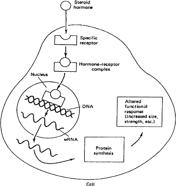
Figure 7.1-an anabolic steroid as it enters the cell and initiates protein synthesis
As mentioned previously, steroids are produced naturally within the body and are fat-soluble organic compounds with a tetracyclic framework derived from cholesterol.
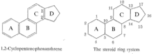
The base structure of steroids is shown below:
There are three major classes of steroids, which are all produced in the adrenal cortex; glucocorticoids, mineralocorticoids, and androgens (although the androgens are mainly synthesized in the gonads-especially the testes and ovaries). As androgens is the class which influences muscle growth the most, these types of steroids (such as testosterone-a key steroid which possesses both anabolic and androgenic properties) will primarily be focused on.
The complicated process in which cholesterol is converted to androgens (androstenedione and dehydroepiandrosterone) and then testosterone is illustrated in Figure 7.3. Firstly, cholesterol is modified by the cytochrome P-450 enzyme, resulting in pregnenolone (figure 7.2). Pregnenolone is essentially the originator of androgens.
Through the use of the 17-hydroxysteroid dehydrogenase enzyme in the testes and ovaries, the androgens can be converted to testosterone.
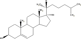
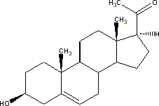
Figure 7.2-line structures for cholesterol and pregnenolone, respectively
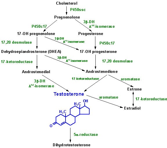
Figure 7.3-eventual conversion from cholesterol to testosterone
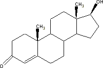
Figure 7.4-structure of testosterone
The most common prohormones (synthetically produced chemicals which are taken manually) are typically converted within the body into anabolic steroids such as testosterone, dihydrotestosterone (DHT), nandrolone (nortestosterone), and 1- testosterone, all of which will be discussed subsequently. Some of the more familiar prohormones advertised for are 4-androstenedione, 4-androstenediol (4-AD), 19- norandrostenedione, 19-norandrostenediol, and 1-androstenediol (1-AD). Because these
are converted to steroids within the body and are not steroids as such themselves, they are legal in the U.S. without perscription until Janurary 19th, 2005 when laws banning prohormones will be implemented.
Within the body, testosterone can be converted to dihydrotestosterone (the most effective of male steroids) by the 5α-reductase enzyme. What occurs is the double bond between C4 and C5 is downgraded to a single bond, thus allowing room for an additional hydrogen atom to bond to the two carbon atoms. As shown in Figure 7.5, the new hydrogen atom on C5 is trans to the methyl group while the new hydrogen atom on the C4 can be either cis or trans due to the already present hydrogen atom.
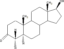
Figure 7.5-structure of dihydrotestosterone
Nandrolone is very similar in structure to testosterone. Following the reaction, a hydrogen atom has replaced the methyl group in C10. Figure 7.6 shows the structure of nandrolone.
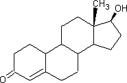
Figure 7.6-structure of nandrolone
1-testosterone (1-dihydrotestosterone) is simply an isomer of testosterone, although it is claimed to be 700% more anabolic and 200% more androgenic than testosterone. The structure of 1-testosterone is illustrated in figure 7.7.
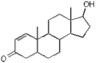
Figure 7.7- structure of 1-testosterone
There are many negative consequences due to taking steroids, although many are reversible upon the termination of steroid intake. Following is a list of possible adverse effects from steroids:
-
- Disruption of hormonal production in the body
- Reduction in sperm cell production and shrinkage of the testicles in men (testicular atrophy)
- Baldness or loss of hair
- Development of breasts in men (gynecomastia)
- Enlargement of the clitoris and decrease of breast size in women
- Coarsening of the skin in women
- Increase in sex drive
- Growth stunt in adolescences
- Injury to tendons, ligaments and/or muscles
- Cardiovascular diseases such as heart attack and stroke
- Disruption of blood flow
- Liver tumour and cysts can form on the liver, possibly rupturing and resulting in internal bleeding
- Acne and cysts
- HIV and hepatitis due to sharing and/or non-sterile needles
- Psychological effects can include rage and delusions
The reason for gynecomastia in men is due to the conversion of testosterone (or another androgenic steroid) to an estrogen (the primary female sex hormone) through a reaction called aromatization (because ring ‘A’ becomes an aromatic ring), where the enzyme aromatase acts as a catalyst. For example, testosterone can be converted in to an estrogen called estradiol, a type of estrogen.
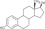
Figure 7.8- structure of the estrogen, estradiol
Below is a table illustrating the characteristics of the prohormones mentioned earlier. Note that conversion rate signifies the amount of the steroid that is converted to testosterone.
Table 7.1- characteristics of prohormones1
| Prohormone | Converts to | Characteristics |
| 4-androstenediol (4-AD) | testosterone |
prostate. |
| 4-androstenediol (4-AD) | testosterone |
(DHT). |
| 19-norandrostenedione | nortestosterone |
|
| 19-norandrostenediol | nortestosterone | Same as -dione, except (as with the andros),
the conversion rate is higher. |
| 1-androstenediol (1-AD) | 1-testosterone
(1-Dihydrotestosterone) |
|
| with excessive levels of DHT, including male pattern baldness, testicular shrinkage, benign prostate hypertrophy and acne can occur with 1-AD usage. (Journal of Organic chem. vol, 27 1962 iss.1) |
Because steroid abuse is a developing issue in the athletic community, testing for steroids have become mandated in events such as the Olympics. To detect the presence of steroids within the body, a urine sample is collected from a human, which is then analyzed by a gas chromatograph and mass spectrometer (GC/MS). A chromatograph is essentially device which is used in combination with a mass spectrometer and is used to separate mixtures before the sample is analyzed by the mass spectrometer. A mass spectrometer is a device which allows the identification of a (unknown) compound based on its mass.
(mass spectrometer)
Figure 7.9 – a gas chromatograph. The injector is heated resulting in the inputted chemical becoming a gas. In the column, the mixture is then separated due to their volatility and the separated chemicals are carried through the tube by an inert gas. Less volatile chemicals will travel with a greater speed than those with more volatility. This diagram is copyrighted by the Oregon State University.
gas chromatograph
Figure 7.10 – a mass spectrometer. Once the mixture has been separated and have entered the mass spectrometer, the chemicals are bombarded with electrons by the ionization source, causes the molecules to form radical cations and subsequently break into smaller pieces. The cations are then accelerated and then pass through a magnetic plate and are deflected. The less the mass is, the more deflection occurs (see Figure 7.11). This diagram is copyrighted by the Oregon State University.
Figure 7.11 – deflection of ions in a mass spectrometer
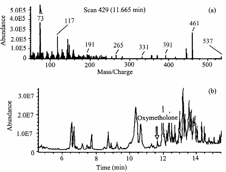
Figure 7.12 -mass spectrum (a) and total ion chromatogram (b) of oxymetholone, an anabolic steroid.
Conclusion
The central idea of this paper is to research various aspects of bodybuilding. Firstly, it was determined that the tearing of muscles due to contractions results in increased protein synthesis which ultimately results in muscle growth. Thus, anaerobic exercise is more effective for muscular growth because powerful contractions occur.
Aerobic exercise results in more muscular endurance, due to the increased efficiency of cellular respiration. Essentially, we can determine the effectiveness of a particular anaerobic workout by calculating the amount of work performed (the intensity of the workout must be adequate, thus a heavy load balanced with a reasonable amount of reputations (e.g. 7-10) results in maximal success). Conceptions that fast reputations are more effective are actually false. This is primarily due to the concept of impulse and the fact that the work done is the same. Furthermore, a faster workout could result in the injury of tendons.
Essentially, the arm of a human being acts as a third class lever; that is to say that a small contraction of the bicep results in a large movement of the load although the bicep is exerting a proportionally larger force. Thus, this further demonstrates the effectiveness of anaerobic exercise resulting in larger muscles.
The idea of promoting an appealing self-image has resulted in the increased demand for supplements and prohormones. Protein and creatine supplements have been especially used for the purpose of increased effectiveness of weight training. Although creatine and protein are ingested through various foods that are consumed daily (such as red meat), although fat and cholestrol are also in these food products and can hinder the process of developing a leaner image. Protein supplement intake results in the increased availability of amino acids which are ultimately responsible for muscle growth due to increased actin and myosin synthesis. Creatine is a source of adenosine triphosphate (ATP) production; ATP is the energy “currency” in the body. Essentially, by ingesting creatine, it is brought in to the bloodstream, then phosphorylated by the creatine kinase and eventually the phosphate group is broken off the phosphocreatine, where the phosphate group is then added to the adenosine diphosphate (ADP), yielding ATP. As well, creatine is advertised to “buffer” the lactic acid (a product of anaerobic respiration) in muscles. In reality, it is not the creatine which buffers lactic acid, as demonstrated by by the lab, although the lab was inconclusive in regard to phosphocreatine. However it is likely that the phosphate acts as the buffer for lactic acid. Phosphate ion acts as a buffer in the intracellular environment. Some athletes load on carbohydrates with the idea that more energy will be produced due to the synthesis of ATP from the breakdown of glucose.
The use of anabolic-androgenic steroids essentially promotes masculine characteristics, such as relatively larger muscles (whereas estrogens promote feminine characteristics). Anabolic steroids are illegal to buy or intake unless they are clinically prescribed. However, prohormones (synthetically produced chemicals which are taken manually are typically converted within the body into anabolic steroids) were legal to
purchase and use in the United States until recently. The side effects of anabolic steroids are quite dangerous, and there are many. For example, men can grow breasts, shrinking of the testicles, stimulation of hair growth, etcetera. Because the intake of steroids are becoming more and more common in the athletic community and thus the process of detecting steroids has become more vigorous and more routine. To detect the presence of anabolic steroids in the human body, urine samples are passed through a mass spectrometer.
Works Cited
Note that there were several sources of information that were used for various different sections, such as Encarta. The most used source for this paper is the tremendously useful textbook, Human Physiology; from cells to systems, 5th edition by Lauralee Sherwood.
Another tremendous help has been from the numerous articles and publications of Richard B. Kreider, Ph. D. As well, note that some of the sources listed may not have been for factual research but instead were used to gain an understanding of the supplement market and advertisement claims. Please recognize that some of the diagrams and photos used in this paper may be copyrighted by the original author. If there are any concerns regarding copyrighted material, the necessary corrections will be made. All resources were retrieved between November 2004 and January 30th, 2005.